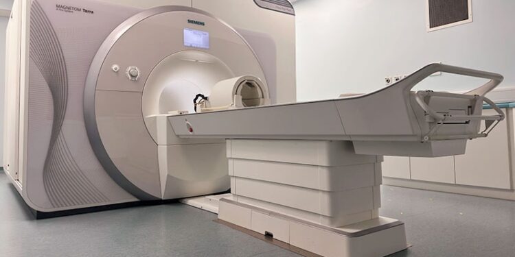
A new MRI technique developed at the University of Cambridge, Cambridge, England, could improve the detection of brain lesions responsible for focal epilepsy. The new method offers greater precision than standard MRI scans, improving the chances of successful surgery.
MRI plays a critical role in identifying structural lesions to aid pre-surgical planning in patients with drug-resistant epilepsy. The chances of being seizure-free after epilepsy surgery are roughly doubled if a lesion is detected with MRI. Recent advancements have focused on ultra-high field 7 Tesla (7T) MRI, which offers superior spatial resolution and sensitivity compared with standard 3 Tesla (3T) MRI. This allows identification of structural epileptogenic lesions that are not detectable using 3T MRI.

Addressing Imaging Challenges in the Temporal Lobes
However, 7T MRI can suffer from signal dropouts in areas with a weak transmit B1+ field. Furthermore, these dropouts often occur in the temporal lobes, which are regions of particular interest for epileptogenic lesion detection. Parallel transmit (pTx) MRI can substantially counteract these dropouts, improving the uniformity of 7T MRI images and ensuring diagnostic quality across the entire brain.
Previously, the use of pTx was limited by the need for time-consuming patient-specific calibration scans. To address this, researchers tested a new “plug and play” system using template-based universal pulses. They compared pTx 7T MRI with standard single-transmit 7T MRI in 31 consecutive adult candidates for epilepsy surgery whose 3T scans were negative or inconclusive.
Improved Visualisation and Clinical Impact
The findings, published in the journal Epilepsia, showed that pTx 7T MRI improved lesion visibility in 57% of cases. The standard 7T MRI never produced better visualisation. Previously unseen structural lesions were identified in nine patients (29%), while four patients (13%) had lesions confirmed, and another four had suspected lesions ruled out. The new technique altered clinical management in 18 cases (58%).
The researchers noted that their new technique “allows much of the benefit of pTx at clinical timescales, with zero time penalty for the user and with no special expertise required.” They recommended that future clinical implementations of 7T MRI for epilepsy “should utilise parallel transmit where possible.”
Expert Perspectives on the Findings
John Duncan, professor of neurology at UCL Queen Square Institute of Neurology, London, England, who was not involved with the study, highlighted the significance of accurate lesion identification for epilepsy surgery. However, he explained to Medscape News UK that currently available clinical MRI scans fail to show a relevant abnormal area in 1 in 3 patients with epilepsy who might benefit from surgical treatment.
Current MRI scanners have powerful magnets with a strength of 3T. Scanners with stronger 7T magnets can identify some subtle abnormalities that are not found on a conventional MRI scan, Duncan noted. “These more powerful scanners, however, may give rise to shadows that make the images hard to interpret.” The Cambridge group developed a novel way of using a 7T MRI scanner, “resulting in images that were clearer and easier to interpret.” This will aid the decision-making for surgical treatment, he said.
Professor Ley Sander, medical director of the Epilepsy Society, welcomed the findings. “We are delighted to learn that by using multiple transmitters, people have had successful surgery to remove the relevant lesions causing their seizures,” he told Medscape News UK. “This underpins the need to fund further research and the benefit of working collaboratively and across disciplines,” he emphasised. Sander stressed the importance of clinical and research staff having access to new technical innovations so they can further develop and pioneer new treatments and approaches.
Tom Shillito, Health Improvement and Research Manager at Epilepsy Action, described the results as promising. He noted that while the technique has potential, widespread adoption may take time. “We are looking forward to seeing more about the potential application of this [technique] and further research in this area,” he told Medscape News UK.
The study was funded by the National Institute for Health and Care Research, the Medical Research Council, and Cambridge University Hospitals Academic Fund.
Sheena Meredith is an established medical writer, editor, and consultant in healthcare communications, with extensive experience writing for medical professionals and the general public. She is qualified in medicine and in law and medical ethics.
Source link : https://www.medscape.com/viewarticle/advanced-mri-technique-improves-diagnosis-drug-resistant-2025a10006ww?src=rss
Author :
Publish date : 2025-03-24 09:13:00
Copyright for syndicated content belongs to the linked Source.


