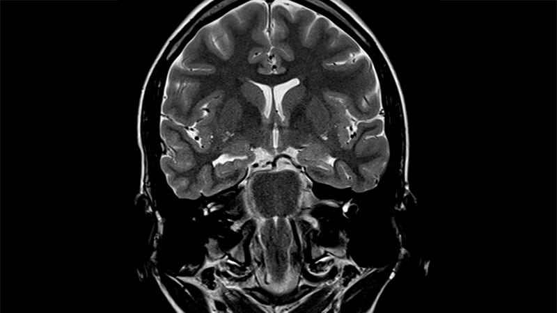
LOS ANGELES — An open-source artificial intelligence (AI) module can help identify epileptic lesions on MRI missed by human reviewers, including both focal cortical dysplasia (FCD) and hippocampal sclerosis, according to new research.
An estimated 42%-55% of adult and pediatric patients with epilepsy who undergo surgery with negative MRI findings have FCDs, which are malformations of cortical development, according to Sophie Adler, MBPhD, who spoke at the American Epilepsy Society (AES) 78th Annual Meeting 2024.
Various efforts have been made to use automated machine learning to identify FCDs. Adler, a medical student at University College London, London, England, and colleagues developed an open-source machine learning program, published in 2022, which uses a neural network for FCD detection based on 33 surface-based features in 618 patients with epilepsy and 397 controls from 22 worldwide epilepsy centers. They trained and cross-validated the network on half of the cohort and then tested it on the other half, in which it had a sensitivity of 59% and a specificity of 54%. The specificity was raised to 67% after the inclusion of a border around lesions that accounted for uncertainty around the borders of manually delineated lesion masks.
Adler noted that there was sparse representation of lower-income countries. “That’s something that we really need to think about addressing and think about when we interpret our results, that these methods have been trained on data from certain centers and are not necessarily fully representative of the world,” she said.
The algorithm examines every surface point of the brain and decides whether it resembles a lesion. That led to the discovery of lesions in 63% of MRI-negative patients, but it also led to a significant number of false positives, leading to a great deal of neurologist efforts to review and confirm such findings, and this has been found to be an issue with other AI methods, according to Adler.
To address the issue, the group employed a graph convolutional neural network. “Instead of just seeing every point on the cortical surface and having to make a decision of ‘is this normal or is this abnormal,’ this convolutional neural network can build up an understanding of what the neighbors of that point are, and its neighbors’ neighbors, and use that to build up an understanding of a whole hemisphere, and therefore be able to learn about context in the brain,” said Adler.
The strategy reduced false positives, raising the positive predictive value to 67%. “So if the algorithm finds something, it’s nearly 70% likely that that really is an FCD, and yet we’re still able to find around 65% of the lesions that were missed by humans,” said Adler.
The group is now working with radiologists and surgeons to bring the algorithm into clinical practice and has also collaborated with two surgeons in a clinical trial to use the algorithm to identify suspected lesions in advance of implanting electrodes.
She also emphasized the open-source nature of the software, and the group is conducting workshops to train clinicians and researchers to use it.
The group has also developed an algorithm to detect hippocampal sclerosis, which is responsible for about 10% of lesions missed by MRI, according to Adler. It relies on a similar surface feature approach with normalization approaches, and the group is now working to incorporate both into a single algorithm. “In terms of finding the lesions we cannot see, we now have algorithms to find FCD, hippocampal sclerosis, and we’re moving towards multi-pathology detection,” said Adler.
During the Q&A session after the talk, an audience member asked about the disagreements often seen in the interpretation of clinical imaging and called for better standardization. “That would feed better into some of the big data analysis,” he said. Sara Inati, MD, who spoke during the session about new imaging methods, agreed on the importance of International League Against Epilepsy imaging standards. “I think if people start to follow those and actually do similar protocols across centers, it actually aids in the efforts…where you can then more easily use these machines. But I think you’re also hitting on another important point, which is most radiologists or neurologists certainly are not well equipped to be creating these complex models. I think what [Adler and others] have been trying to do is make this more accessible to us, and as they do the hard work of working out all the technical details, I think the hope is that it will become easier for clinicians to adopt. I think we’re just starting to get there, but I don’t think that [will be] current in 2025,” said Inati, who is an assistant clinical investigator in the NINDS Intramural Research Program.
Inati and Adler had no relevant financial disclosures.
Source link : https://www.medscape.com/viewarticle/ai-catches-missing-mri-lesions-2024a1000p16?src=rss
Author :
Publish date : 2024-12-24 02:53:37
Copyright for syndicated content belongs to the linked Source.
