
[ad_1]
CORONADO, CALIF. — According to Christopher J. Arpey, MD, there is no single best approach to treating lentigo maligna (LM) and LM melanoma (LMM), but Mohs micrographic surgery is a sensible option in cases where margin size is critical and tissue conservation with precision is essential.
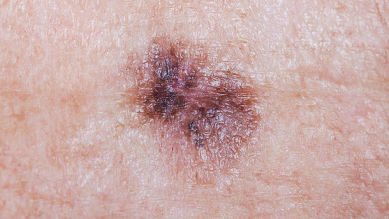
At the annual Melanoma and Cutaneous Oncology Symposium, Arpey, professor and chair of dermatology at Mayo Clinic Arizona, noted that LM and LMM comprise about 10% of all cases of malignant melanoma, with a rising incidence as documented by Surveillance, Epidemiology, and End Results cancer registry data. The estimated risk for progression of LM to LMM is 2%-5%.
“The management of these patients is dependent on a lot of factors,” he said. “What I say to a lot of our trainees is that not every actinic keratosis becomes a basal cell carcinoma or squamous cell carcinoma. Is every LM going to progress to LMM? Maybe not. If LM involves an entire eyelid or other critical anatomic location, or if the person is elderly or has a lot of comorbidities, maybe observation’s okay. But there is clear-cut progression in some cases.”
However, while several modified surgical techniques that use permanent sections exist for the management of melanoma, he continued, there is no gold standard for Mohs surgery for LM/LMM, which adds to the complexity of treatment, as the sizes and location of pigmented lesions vary.
‘No Guarantees’ With Any Treatment
Then there’s the fact that LM can be multifocal, so “clear margins” are relative only. Subtotal initial biopsies might miss the invasive MM. “Explain to the patient that there are no guarantees with any LM treatment,” Arpey advised. “If you are not going to operate, you need baseline photos and periodic surveillance biopsies. Counsel them that not every lentigo is a lentigo maligna.”
For the hypervigilant patient, “I consider adjuvant off-label topical therapy with imiquimod if we’re just going to leave things be, though the data is incomplete,” he said. In 2023, Italian researchers published a systemic review of 87 articles on the topic and found a high variability in response rates and a mixture of protocols, with histological clearance achieved in 56.9% of lesions and clinical clearance achieved in 43.2%.
Radiation therapy is another non-surgical option, he said. In a 2020 systematic review of 14 studies of radiotherapy involving 1,243 LM and LMM lesions, Swiss researchers observed local recurrence rates of 0%-30%.
If he chooses to treat the LM surgically, Arpey said he sends the central portion to pathology to rule out invasion prior to taking the initial Mohs layer to treat the periphery and delays the repair if it requires a flap or graft. “Sometimes, I’ll use 8 mm debulking margins plus 2 mm initial Mohs margins, so that I have sufficient ‘standard’ margins if the path report surprises me,” he said at the meeting, which was hosted by the Scripps Cancer Center, San Diego.
On histology, LM appears as nested or confluent clearly atypical melanocytes with or without pagetosis. “Typically, you need to see at least three of these melanocytes together to call it a nest,” he said. Melanoma-associated antigen recognized by T cells (MART-1) or Melan-A stains can help distinguish melanocytes, particularly if freeze artifact is present, he said. However, these stains do not differentiate between benign and malignant cells. They can also stain melanosomes and melanophages.
“Be careful not to overcall sun-damaged melanocytes, because this can lead to face-ectomies,” Arpey said. “Using ‘control’ samples on the opposite anatomic side may help for comparison of melanocyte density. You have to be diligent about this. One of the biggest things we’re missing as a dermatologic surgical community is standardization of the approach and best practices.”
The Role of Stains
Arpey recommended a two-part CME series published in 2024 in the Journal of the American Academy of Dermatology. Part I covers the epidemiology, risk factors, and diagnosis of LM, while Part II addresses management. In Arpey’s clinical experience, MART-1 is the most common stain used for LM and LMM, along with microphthalmia-associated transcription factor (MiTF) or Sry-related HMG-box-10. He explained that MART-1 is a cytoplasmic stain and stains melanin, while MiTF is a nuclear stain, and stains melanocytes.
“MiTF is more specific, and especially helpful in heavily tanned skin — like in Arizona,” he said. “However, it also stains dermal dendritic cells, some hair root sheaths, and some adnexal structures. MART-1 is better for picking up islands/nests of melanocytes where known microinvasion is in play.” He emphasized that these stains are “are not a panacea, but an adjunct” to the H&E technique. He characterized preferentially expressed antigen in melanoma staining as “a work in progress for predicting proliferative/malignant potential of melanocytes.”
Reported recurrence rates of LM treated with Mohs micrographic surgery have ranged from 0.5% to 30% in the literature, he said, with a time to recurrence typically in the range of 4-7 years. Other modalities such as topical and radiation therapy have higher rates of recurrence compared with margin-controlled surgery.
Many studies have shown 0.5 cm excisional margins for LM to be inadequate, while recent guidelines from the American Academy of Dermatology recommend margins of 5-10 mm. “Treatment of LM with MMS has been shown to lower recurrence rates significantly,” he said. “More recent studies suggest about 60% clear margins with standard excision technique with 6 mm margins and close to 90% at approximately 9 mm.”
Arpey explained that he sends debulked central tissue for histological evaluation because, although melanoma in situ has nearly a 100% 5-year survival rate, any level of invasion worsens the prognosis. Rates of invasion found on debulked specimens initially thought to be LM are thought to be around 5%. A series of 173 cases showed upstaging in 8%.
A larger, more recent study performed at Mayo Clinic found that 4% of LM with subtotal biopsy sampled showed invasion. “Immunostaining and rush paraffin sectioning techniques have been developed in hopes of improving clearance rates,” he said. “However, their use results in increased cost ($30-$100) and procedure time (19-60–minutes-plus),” he said. “It’s an adjunct. It’s not going to give you the answer. It’s a tool to help you with the interpretation.”
Arpey cited two comparative studies, one of 662 cases and one of 359 cases, which demonstrated that mortality is equivalent with wide local excision vs Mohs. However, “the morbidity spared and ability to reconstruct immediately [with Mohs] is a huge advantage,” he said. “Even though we cannot demonstrate superiority for Mohs for melanoma in situ or even lower stage melanoma, morbidity matters. Disease-free survival doesn’t have to be your only endpoint.”
Arpey reported having no relevant disclosures.
[ad_2]
Source link : https://www.medscape.com/viewarticle/expert-tips-mohs-surgery-melanoma-2025a10002ya?src=rss
Author :
Publish date : 2025-02-06 08:31:33
Copyright for syndicated content belongs to the linked Source.