
[ad_1]
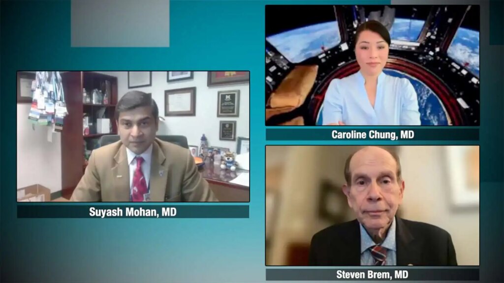
Following the American Society of Neuroradiology meeting, MedPage Today convened three leaders in neuro-oncology and brain tumor imaging for a virtual roundtable discussion on the evolving role of advanced MRI in brain tumor care. Moderator Suyash Mohan, MD, of the University of Pennsylvania in Philadelphia, is joined by Caroline Chung, MD, of the University of Texas MD Anderson Cancer Center in Houston, and Steven Brem, MD, also of the University of Pennsylvania.
In this fourth and final episode, the panel looks ahead to the future of brain tumor imaging, discussing how quantitative imaging biomarkers, artificial intelligence (AI), and public-private collaboration could advance precision neuro-oncology. The experts also consider what evidence will be needed before AI-derived imaging signatures can be trusted to guide real-world clinical decisions.
You can view the full series here.
Following is a transcript of their remarks:
Mohan: Now, since we are coming to the end of this session, I’d like to ask a little bit about the future in terms of quantitative biomarkers and artificial intelligence.
And again, starting with Dr. Chung, looking ahead 3 to 5 years, what do you think is actually going to make it into routine brain tumor practice? What do you think is ready for prime time? What is near horizon? And what do you think still needs more work in terms of validation and accessibility?
Chung: So in terms of where I think this conversation has gone and what we would like to see, being able to, what I call in vivo imaging. Could we actually bring in vivo biological understanding from a noninvasive procedure like a multiparametric MRI? The imaging-pathology correlation are critical steps. And I think with each of these that I’m going to highlight, there are interdependencies.
So if we can actually gain biological understanding from this imaging, this is an art of the possible. I think the technology is there. We need to be able to gather the meaningful data to actually allow that translation to happen. And that would be an incredibly exciting future because anytime we want to gain biological understanding at this point, at this moment in time, for brain tumors at least, it means a brain tumor surgery.
And I think that it’s a necessary step for some patients. It’s something that I think every patient would love to avoid if they can help it. And I think as much as Dr. Brem, I’m sure you’d want to get the meaningful tissue if you could avoid opening someone’s skull up to get that meaningful tissue and get insights, I’m sure we would all want to do what’s best for our patients in terms of minimizing the invasive nature when needed. And we need to save the time in the OR [operating room] to actually get the patients in who really do need that surgical intervention.
In terms of scalability, I think it really depends on, again, it depends on us. It comes down to us collectively working together to say, “This is something we can do uniformly and we can actually leverage this and scale it in a meaningful way when it comes to quantitative imaging.”
There are many different efforts that are happening. Even with the Quantitative Medical Imaging Coalition, we work with our industry vendors. We have industry representation at the table, both from the imaging vendors who are helping us, who are actually building out a lot of these prepackaged analytic programs and prepackaged pulse sequence packages for specific indications.
I think we need to encourage our community and other centers, and collectively with each other, to start to adopt some of these that we can actually more meaningfully cross-calibrate imaging measurements and harmonize them so that we can actually look at longitudinal changes in our patients in a meaningful way. And if we were to do that, the insights that we gain from longitudinal changes would go up by default.
The final piece is that I think that the public-private partnerships and endorsing those pieces is going to be a critical piece. A lot of incredible innovation is happening at academic centers, but to scale that out to the masses we need to have the public-private partnerships to actually bring together packaged educational programs along with packaged delivery programs that can be implemented broadly. And the public-private partnerships then from the actual image acquisition, the image processing and leveraging, perhaps even the AI technologies help us fill the gaps.
To your point, imaging does take time, but there’s a lot of technology that’s emerging to say, can we fill the gaps leveraging technology like AI? So if you look at MR fingerprinting and getting quantitative T1, T2 without necessarily getting the actual acquisitions, can we actually leverage some of that technology? There’s synthetic MR that’s emerging out of CT scanners. This is in the global health space, something that may be a necessary step to bringing forward information that was just not… the technology is just not there physically in terms of the hardware in certain centers. These are all things that we could start to explore in terms of disseminating and generating access to care for those who are not in areas where the technologies are actually available.
Mohan: Yeah, thank you. And along those lines of emerging AI technologies, Dr. Brem, if I can ask you a follow-up question that from a surgeon’s chair, what would make you trust an AI-derived imaging signature enough to act on it for a real patient?
Brem: Well, I think what would make me work on AI, we always have the ground truth. And the nice thing about being a surgeon is you actually go into the operating room and you touch, have all kinds of senses so you know you’re not just looking at an image, you’re really looking at a biologically active tumor and it’s right there.
So I think that when we had the Society for Neuro-Oncology meeting and we spoke with Alex Golby, who’s done a lot of innovative work at the interface of imaging and neurosurgery, she said every neurosurgeon goes into an operating room, the more they know, the more they can do. So we would have to see if all the data coming from the quantitative imaging consortium that Caroline is running, whether that’s really noise or really signal. And as with DTI [diffusion tensor imaging], as with any new tool, you learn where it really helps and really doesn’t.
I wrote a commentary regarding how much modality do we actually need when we do take out a brain tumor? Do we need fluorescence? Do we need DTI? Do we need ultrasound? Do we need, on and on, metabolic mapping? How many maps do you need? And it depends on the center, depends on the surgeon to achieve those goals of maximal safe resection. I think it’s going to be an incredible tool, but we want to avoid the type I errors and type II errors. We don’t want to have false positives and false negatives.
Working on a project with Dr. Ragini Verma now, and by altering the settings, we were looking for the sweet spot with the DTI and edema correction and if it’s too sensitive, too specific. If it’s too sensitive, we get spurious tracks. On the other hand, we don’t want to miss anything. So it’s adaptive, it’s individual. Obviously we’re making enormous progress.
I love that you brought up, Caroline, the AP4. There was a concept from the NCI [National Cancer Institute] at the turn of the century that the future of oncology was going to be academic private-public partnership programs, AP4 programs. And now I believe they have something with ARPA [American Rescue Plan Act] and maybe your consortium will get the kind of funding that you need, but to harness all of the brainpower and to get the proper data sets and to get a large enough database so we can make meaningful comparisons and really filter out what’s the signal, what’s noise.
And all this new technology always goes through the Gartner hope-hype curve where, wow, this is amazing, and then you find out there is some downside. Then you say, well, maybe it’s going to really have specific… you can’t do certain tumors without AI. It just would be ridiculous. So we know there’s a great deal of variation in the WHO [World Health Organization]… well, how many brain tumor types are there? One hundred and twenty or so? GBM [glioblastoma] itself has so many phenotypes.
Mohan: So thank you. I agree. I’ve heard this from you and others that no two glioblastomas are the same. But thank you both for taking the time to speak with me today. I really appreciated your insights and your thoughtful perspective.
Please enable JavaScript to view the comments powered by Disqus.
[ad_2]
Source link : https://www.medpagetoday.com/meetingcoverage/asnrexpertvideoroundtable/122099
Author :
Publish date : 2026-07-08 18:06:00
Copyright for syndicated content belongs to the linked Source.