TOPLINE:
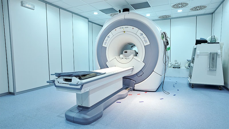
According to a UK study, whole-body MRI showed that patients with arthralgia who had previously received immune checkpoint inhibitor therapy had similar levels of inflammation and erosions as those with clinical arthritis, with distinct imaging patterns.
METHODOLOGY:
- Researchers recruited 60 adults (mean age, 65 years; 43% women; 100% White) with new musculoskeletal symptoms during or within 6 months after receiving treatment with immune checkpoint inhibitors and 20 healthy control individuals (mean age, 62 years; 40% women; 100% White) from the UK, who underwent gadolinium contrast-enhanced whole-body MRI.
- A total of 35 patients had arthralgia and 25 had inflammatory arthritis.
- Two independent masked assessors graded joint, tendon, bursal, entheseal, and whole spinal imaging lesions, with consensus reporting and a 6-month follow-up.
TAKEAWAY:
- Distinct global inflammatory patterns were identified on the MRI scans: polymyalgia rheumatica (extracapsular; 12%), peripheral inflammatory arthritis (37%), spondyloarthropathy (n = 1), and a non-specific pattern (20%).
- Median total appendicular joint synovitis, joint erosion, enthesitis, and tenosynovitis scores were significantly higher in both arthralgia and inflammatory arthritis groups than in control individuals, with no significant differences found between the arthralgia and inflammatory arthritis subgroups.
- Among the patients exposed to immune checkpoint inhibitors, acromioclavicular (77%), glenohumeral (75%), wrist (73%), and metacarpophalangeal (59%) joints were most frequently affected by synovitis.
- Patients with a peripheral inflammatory arthritis pattern were most likely to require disease-modifying antirheumatic drug therapy, showing the highest initial and ongoing glucocorticoid requirement.
IN PRACTICE:
“[The study] results suggest that the overall burden of musculoskeletal toxicity associated with immune checkpoint inhibitors is currently under-recognised and highlights the range and extent of inflammation in all patients presenting with new musculoskeletal symptoms after exposure to an immune checkpoint inhibitor. Rheumatology assessment should be considered by oncologists for patients who develop arthralgia alone,” the authors wrote.
SOURCE:
This study was led by Kate Harnden, MBChB, Leeds Institute of Rheumatic and Musculoskeletal Medicine, Leeds, England. It was published online on June 10, 2025, in The Lancet Rheumatology.
LIMITATIONS:
Feet and ankles were not imaged during the study due to logistical constraints. The 6-month follow-up period was relatively short for determining long-term musculoskeletal outcomes. Some patients were classified as having non-specific inflammation patterns, whose significance was unclear. A few participants had preexisting autoimmune diseases, possibly increasing their baseline risk for arthritis. Additionally, intra-assessor agreement was not measured.
DISCLOSURES:
This study received support from the National Institute for Health and Care Research Leeds Biomedical Research Centre. One author reported receiving a EULAR bursary travel award, and some authors reported having financial ties with various sources. Details are provided in the original article.
This article was created using several editorial tools, including AI, as part of the process. Human editors reviewed this content before publication.
Source link : https://www.medscape.com/viewarticle/mri-reveals-joint-damage-after-cancer-treatment-2025a1000h12?src=rss
Author :
Publish date : 2025-06-27 12:00:00
Copyright for syndicated content belongs to the linked Source.
