TOPLINE:
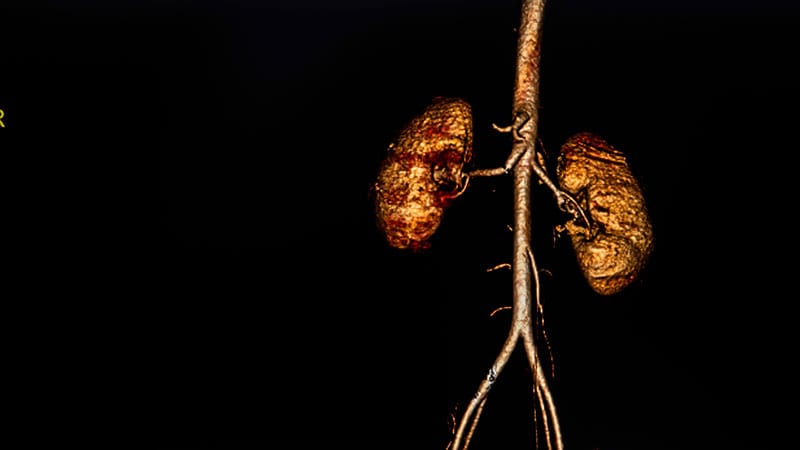
Compared with energy-integrating CT (EICT), ultrahigh-resolution (UHR) photon-counting CT (PCCT) enhanced the visualisation of small vascular structures in aortic imaging while reducing radiation exposure, a study showed.
METHODOLOGY:
- This retrospective analysis included 50 patients divided into two groups: 25 patients (mean age, 65.0 years; 16 men) underwent UHR abdominal CT angiography using PCCT and 25 matched control individuals (mean age, 69.8 years; 17 men) underwent conventional EICT.
- The signal-to-noise ratio (SNR) and contrast-to-noise ratio (CNR) were measured, and image quality was evaluated using a 5-point Likert scale.
- Radiation exposure was compared using volume CT dose index (CTDIvol), dose length product, and effective dose measurements.
TAKEAWAY:
- Radiation exposure was significantly lower in the PCCT group than in the EICT group, with a CTDIvol of 4.7 mGy vs 7.3 mGy (P = .0081) and an effective dose of 3.4 mSv vs 6.5 mSv (P = .0057).
- The analysis revealed a higher SNR and CNR in PCCT than in EICT, though statistical significance was achieved only for renal arteries (P = .0432).
- Subjective image quality was significantly better with PCCT than with EICT (P < .0001).
- Analysis of visual grading characteristics showed excellent diagnostic accuracy with an area under the curve of 0.9 for renal arteries, the inferior mesenteric artery, and lumbar arteries.
IN PRACTICE:
“As the implementation of the UHR mode did not result in higher radiation dose, nor did it lead to a significant reduction of CNR in comparison to EICT, the additionally available improvement in assessing small vessels should lead to further use of UHR mode in clinical routine,” the authors wrote.
SOURCE:
The study was led by Isabelle Ayx, MD, University Medical Center Mannheim, Heidelberg University, Mannheim, Germany. It was published online on March 13, 2025, in the European Journal of Radiology.
LIMITATIONS:
The study was limited by the small sample size and the use of matched control individuals instead of the same patient population, which reduced the strength of the comparison analysis. The retrospective setting limited the collection of complete demographic data, including height, weight, and body mass index. The image quality evaluation, although conducted by two radiologists blinded to scanner types, may have carried a potential bias toward the new method.
DISCLOSURES:
The study was funded by Gesundheitsstandort Baden-Württemberg. The authors reported having no conflicts of interest.
This article was created using several editorial tools, including AI, as part of the process. Human editors reviewed this content before publication.
Source link : https://www.medscape.com/viewarticle/photon-counting-ct-lowers-dose-improves-vascular-imaging-2025a10006jm?src=rss
Author :
Publish date : 2025-03-21 12:00:00
Copyright for syndicated content belongs to the linked Source.


