
[ad_1]
Key Takeaways
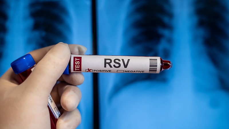
This report describes the case of a 25-year-old woman who presented to the emergency department of the University Hospital St. John Medical Center in Westlake, Ohio, with a syncopal episode. The repeated loss of consciousness was due to acute blood loss resulting from warm autoimmune haemolytic anaemia (AIHA). AIHA can be primary or secondary to an underlying disease that compromises the immune system. In this case, respiratory syncytial virus (RSV) infection was identified as a rare secondary cause of AIHA.
The Patient and Her History
The patient presented with a syncopal episode. She reported a 4-day history of nonproductive cough, nasal congestion, odynophagia, chest pain, shortness of breath, abdominal pain, nausea, vomiting, subjective fever, and headache. She had a history of asthma and depression but was not on anticoagulant treatment and denied any history of trauma or gastrointestinal bleeding.
During the initial evaluation, she experienced another syncopal episode and fell from the treatment table. On arrival, the patient was febrile (39.9 °C), pale, tachycardic (110 beats/min), with a normal respiratory rate (16 breaths/min), blood pressure of 132/68 mm Hg, and oxygen saturation of 100% on room air. Physical examination revealed a cephalohematoma.
Given her abnormal vital signs and recurrent syncope, the following investigations were performed:
- ECG
- Complete blood cell count with automatic differential diagnosis
- Comprehensive metabolic panel
- Exclusion of venous thromboembolism
- D-dimer
- Troponin
- Lactate
- Human chorionic gonadotropin
- Urinalysis
- Viral testing (influenza A/B, severe acute respiratory syndrome coronavirus 2, and RSV)
- Blood cultures
- Chest x-ray
- CT angiography of the chest to rule out pulmonary embolism
She received 650 mg of paracetamol for fever and a 1000 mL bolus of Ringer’s lactate for tachycardia and syncope.
The patient was also administered ipratropium-albuterol nebulised solution with 125 mg of methylprednisolone sodium succinate to treat her asthma exacerbation.
Findings and Diagnosis
ECG showed sinus tachycardia (116 beats/min) with no ischaemic changes or injury patterns, a normal QT interval, no delta wave, no signs of Brugada syndrome, or conduction blocks. Polymerase chain reaction analysis confirmed RSV infection.
The blood cell count showed normocytic anaemia (haemoglobin, 7.3 g/dL), no leucocytosis, and a normal platelet count. A comprehensive metabolic panel revealed elevated total bilirubin (3.6 mg/dL). The D-dimer level was mildly elevated at 501 ng/mL. Liver and renal function tests were normal.
CT angiography of the chest was negative for any evidence of pulmonary embolism but showed patchy, multifocal ground glass and semisolid centrilobular nodular opacities in the left upper and lower lungs, suggestive of bronchopneumonia with bronchiolitis.
In the setting of new anaemia and elevated bilirubin levels, there was suspicion of haemolysis. Therefore, reticulocytes, lactate dehydrogenase, haptoglobin, and fractionated bilirubin were included in the laboratory evaluation. Direct bilirubin resulted at 0.5 mg/dL, and indirect bilirubin was elevated at 3.1 mg/dL. The reticulocyte count was elevated at 7.6%, and lactate dehydrogenase was elevated at 436 U/L, both consistent with haemolytic anaemia.
Given the new onset of anaemia and syncope, she received a transfusion of 1 unit of packed red blood cells. The haematologist recommended hospital admission and intravenous steroids.
Management
Despite the transfusion being delayed, her haemoglobin level dropped to 6.1 g/dL on day 1 of hospitalisation. She continued receiving 150 mg of intravenous methylprednisolone and was started on ceftriaxone and azithromycin to treat the bronchopneumonia noted on her chest CT scan.
The Haematology Department ordered direct antiglobulin test (DAT), DAT immunoglobulin G (IgG), and DAT complement. The DAT was positive, DAT IgG was positive, and DAT complement was negative, all of which were consistent with warm haemolytic anaemia.
On day 2 of hospitalisation, her haemoglobin level dropped further to 5.7 g/dL, necessitating an additional 2 units of red blood cell transfusions. On day 3 of hospitalisation, the haemoglobin level remained stable at 8.1 g/dL.
Because the haemoglobin level remained stable on day 3, she was prescribed 40 mg of oral prednisone daily for 1 week. The patient was discharged on day 3 of hospitalisation with an outpatient haematology follow-up. There, she received a blood transfusion and intravenous rituximab but did not tolerate the infusion. Four weeks post-discharge, her haemoglobin level remained stable, with no further signs of haemolysis.
Discussion
AIHA is commonly triggered by viral infections, such as Epstein-Barr virus. However, RSV has rarely been reported as a secondary cause of haemolysis. Most documented cases of RSV-associated haemolytic anaemia involve paroxysmal cold haemoglobinuria, while this case showed warm AIHA.
AIHA is caused by the destruction of red blood cells by IgG autoantibodies. The main treatment methods are steroids, splenectomy, and immunosuppressants, with rituximab being used in steroid-refractory cases.
Syncope is a common complaint in the emergency department and is often secondary to a benign cause. A thorough medical history helps assess a patient’s risk, with complete evaluation including physical examination, laboratory and radiologic testing, and occasionally hospital admission for further management.
This article was translated from Univadis Germany using several editorial tools, including AI, as part of the process. Human editors reviewed this content before publication.
[ad_2]
Source link : https://www.medscape.com/viewarticle/rsv-triggers-autoimmune-haemolytic-anaemia-young-woman-2025a10004rw?src=rss
Author :
Publish date : 2025-02-25 10:08:07
Copyright for syndicated content belongs to the linked Source.