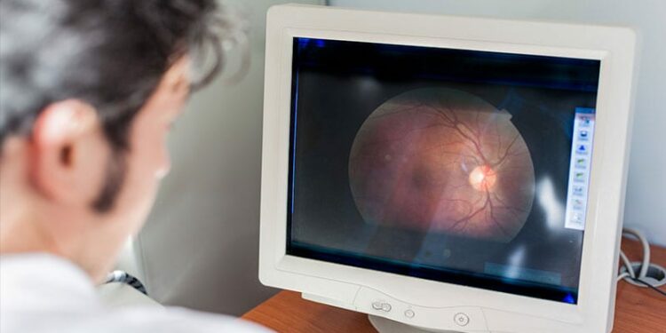
Researchers have identified novel retinal vascular signatures that are significantly associated with the risk for a first stroke. When combined with age and sex, these retinal “fingerprints” predicted stroke risk as well as established traditional risk factors.
The fingerprint, which consists of 29 retinal parameters that measure retinal health, can be detected through routine fundus imaging scans of the back of the eye, investigators said.
“Given that age and sex are readily available, and retinal parameters can be obtained through routine fundus photography, this model presents a practical and easily implementable approach for incident stroke risk assessment, particularly for primary healthcare and low-resource settings,” the study authors wrote.
The findings were published online on January 13 in the journal Heart.
Top Predictors
The retina’s intricate vascular network shares common anatomical and physiological features with the vasculature of the brain, but its potential for stroke risk prediction has not been fully explored.
In the current study, researchers used an advanced artificial intelligence algorithm — retina-based microvascular health assessment system — to analyze 118 retinal vascular parameters using fundus photographs from 45,161 participants in the UK Biobank cohort.
During a median follow-up of 12.5 years, 749 incident strokes occurred. Compared with participants who didn’t have a stroke, those who did were significantly older, more likely to be men, current smokers, and have diabetes. They also had higher body mass index and blood pressure and lower high-density lipoprotein cholesterol.
After adjusting for traditional risk factors, 29 retinal vascular parameters were strongly linked to a first stroke.
More than half (17) were related to density, highlighting the unique role of retinal density in assessing incident stroke risk, reported Mayinuer Yusufu, MBBS, with the Centre for Eye Research Australia, The Royal Victorian Eye and Ear Hospital, East Melbourne, Australia, and colleagues.
Each SD increase in the arc length in arteries, vessels within the macular region, and terminal and non-terminal arteries was associated with an increased risk for stroke, ranging from 9.8% to 14.4%.
Decreases in other identified density parameters were all associated with increased stroke risk, the study team noted.
Specifically, one SD decrease in arterial bifurcation density increased risk by 10.7%, and one SD decrease in branching density in arteries, veins, and vessels in the macula increased risk by 9.9%-12.1%. Each SD decrease in the identified parameters of vessel area density and vessel skeleton density increased the risk for stroke by 11.6%-19.0%.
Among the three identified calibre parameters, central retinal artery equivalent (CRAE), and parameters of length diameter ratio (LDR) were significantly associated with the risk for first stroke in the fully adjusted model.
Each SD decrease in CRAE was associated with a 10.4% increase in stroke risk, whereas each SD increase in arterial LDR and LDR of vessels in the macula increased the risk for stroke by 14.1% and 10.1%, respectively.
For the eight identified retinal complexity measures and the one identified tortuosity measure, each SD decrease was linked to increased stroke risk, ranging from 10.4$ to 19.5%.
Adding these retinal vascular parameters to traditional risk factors improved the area under the receiver operating characteristic curve (AUC) from 0.739 to 0.752 (P
“This is only a small increase in AUC and likely would have little impact on screening performance. That said, when using age, sex and retinal vascular parameters, the AUC was similar to the fully adjusted traditional model (0.739 vs 0.738),” the authors noted.
“Further investigation of these parameters may provide valuable insights into the intricate pathophysiological processes associated with stroke, thereby contributing to the refinement of preventive and therapeutic strategies,” they concluded.
A Promising Development
Commenting on this research for Medscape Medical News, Pierre Fayad, MD, professor of Department of Neurological Sciences, and chief of Division of Vascular Neurology and Stroke, University of Nebraska Medical Center, Omaha, Nebraska, said this study “advances further the knowledge about relationship or retinal vascular changes and risk of stroke, by identifying several new potential measurements that correlate with risk of stroke.”
“This is a promising development. It could easily become automated and routine and will encourage routine and regular eye examinations. However, this still needs prospective testing to validate the findings,” said Fayad, who wasn’t involved in the study.
This research had no commercial funding. Yusufu and Fayad had no relevant conflicts of interest.
Source link : https://www.medscape.com/viewarticle/simple-retinal-scan-may-help-predict-stroke-risk-2025a10001t3?src=rss
Author :
Publish date : 2025-01-24 11:37:31
Copyright for syndicated content belongs to the linked Source.


