In patients with gastroesophageal reflux (GERD) symptoms undergoing screening upper endoscopy, adjunctive use of wide-area transepithelial sampling with 3D computer-assisted analysis (WATS-3D) increases detection of Barrett’s esophagus (BE) and dysplasia, new research showed.
Compared with forceps biopsies (FB) alone, the addition of WATS-3D led to confirmation of BE in an additional one fifth of patients, roughly doubled dysplasia diagnoses, and influenced clinical management in the majority of patients.
“The big take-home point here is that the use of WATS-3D brushing along with conventional biopsies increases the likelihood that intestinal metaplasia will be identified,” first author Nicholas Shaheen, MD, MPH, with the Center for Esophageal Diseases and Swallowing, University of North Carolina School of Medicine at Chapel Hill, North Carolina, told Medscape Medical News.
“Almost 20% of patients who harbor BE were only identified by WATS-3D and might have otherwise gone undiagnosed had only forceps biopsies been performed,” Shaheen said.
The study is published in the American Journal of Gastroenterology.
Beyond Traditional Biopsies
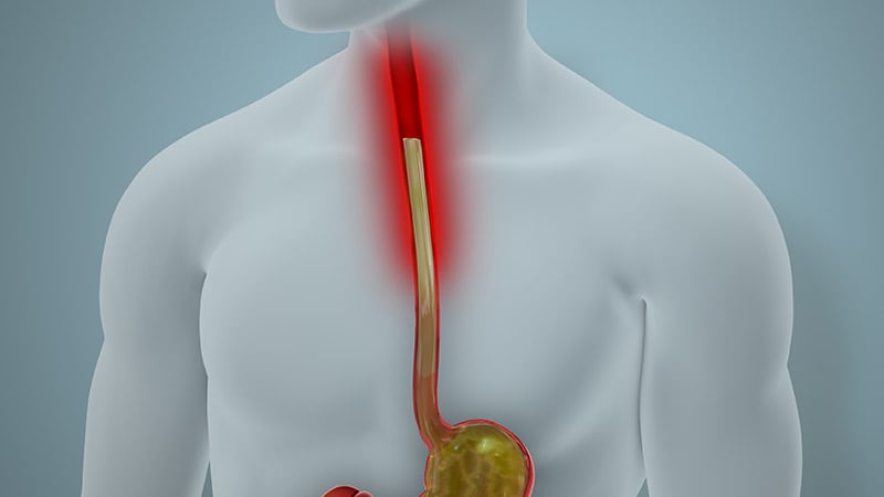
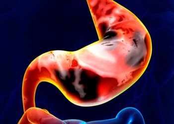
BE develops as a complication of chronic GERD and is the chief precursor to esophageal adenocarcinoma. Early detection of BE and dysplasia is crucial to enable timely intervention.
The current gold standard for BE screening involves upper endoscopy with FB following the Seattle protocol, which consists of four-quadrant biopsies from every 1-2 cm of areas of columnar-lined epithelium (CLE) to confirm the presence of intestinal metaplasia. However, this protocol is prone to sampling errors and high false-negative rates, leading to repeat endoscopy, the study team pointed out.
WATS-3D (CDx Diagnostics) is a complementary technique designed to improve diagnostic yield by using brush biopsy to sample more tissue than routine biopsies.
WATS-3D has been shown to increase detection of dysplasia in patients with BE undergoing surveillance for BE, but less is known about the value of WATS-3D for BE screening in a community-based cohort of patients with GERD.
To investigate, Shaheen and colleagues studied 23,933 consecutive patients enrolled in a prospective observational registry assessing the utility of WATS-3D in the screening of symptomatic GERD patients for BE.
Patients had both WATS-3D and FB in the same endoscopic session. No patient had a history of BE, intestinal metaplasia or dysplasia in esophageal mucosa, or esophageal surgery, endoscopic ablation or endoscopic mucosal resection prior to enrollment.
Overall, 6829 patients (29%) met endoscopic criteria for BE (≥ 1 cm esophageal CLE with accompanying biopsies showing intestinal metaplasia).
Of these, 2878 (42%) had intestinal metaplasia identified by either FB or WATS-3D, but 19.3% had their BE diagnosis confirmed solely on the basis of WATS-3D findings.
Among patients who fulfilled the endoscopic criteria for BE, the adjunctive yield of WATS-3D was 76.5% and the absolute yield was 18.1%.
Of the 240 (1.0%) patients with dysplasia, 107 (45%) were found solely by WATS-3D.
‘Clinically Valuable Adjunct’
Among patients with positive WATS-3D but negative FB results, clinical management changed in 90.7% of cases, mostly involving initiation or modification of surveillance and proton pump inhibitor therapy.
These results suggest that WATS-3D is a “clinically valuable adjunct” to FB for the diagnosis of BE when used as a screening tool in symptomatic GERD patients and particularly in patients with endoscopic evidence of > 1 cm esophageal columnar-lined epithelium, the study team wrote.
Adjunctive use of WATS-3D when BE is suspected “may save endoscopies and lead to quicker, more accurate diagnoses,” they added.
The investigators said a limitation of the study is the lack of central pathology review, potentially leading to diagnostic variability. They also noted that over half of the detected dysplasia cases were crypt dysplasia or indefinite for dysplasia, raising concerns about clinical significance.
Reached for comment, Philip O. Katz, MD, professor of medicine and director of the GI Function Laboratories, Weill Cornell Medicine in New York, said he’s been using WATS for more than a decade as an adjunct to standard biopsy in patients undergoing screening and surveillance for BE and finds it clinically helpful in managing his patients.
This new study provides “further information that WATS added to biopsy that has been traditionally done with the Seattle protocol increases the yield of intestinal metaplasia and likely dysplasia in patients being screened for Barrett’s,” Katz, who wasn’t involved in the study, told Medscape Medical News.
Funding for the study was provided by CDx Diagnostics. Shaheen and several coauthors disclosed relationships with the company. Katz disclosed relationships (consultant/advisor) for Phathom Pharmaceuticals and Sebella.
Source link : https://www.medscape.com/viewarticle/wats-3d-biopsy-increases-detection-barretts-esophagus-gerd-2025a1000711?src=rss
Author :
Publish date : 2025-03-25 10:21:00
Copyright for syndicated content belongs to the linked Source.


