- In advanced heart failure, cardiac remuscularization with BioVAT was associated with an increase in the target heart wall thickness, left ventricular ejection fraction, and quality of life in a small early-stage study.
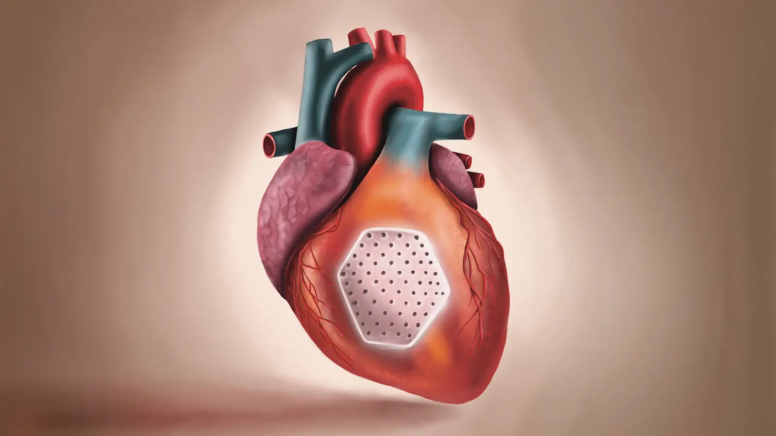
- BioVAT consists of thin patches of engineered cardiac muscle that can be surgically attached to the outside of the ventricle where cardiomyocytes have been lost.
- Questions of efficacy and potential electrical dysfunction require longer-term follow-up and further clinical investigation.
For people with advanced heart failure, modular tissue-engineered patches showed promise as a way to remuscularize the heart, according to interim data.
Biologic ventricular assist tissue (BioVAT) was epicardially transplanted in 20 patients with symptomatic heart failure with reduced left ventricular ejection fraction (LVEF) related to ischemic cardiomyopathy, reported Wolfram-Hubertus Zimmermann, MD, of University Medical Center Göttingen-Georg August University in Germany.
For the 16 individuals treated with the safe maximal dose of BioVAT (20 engineered-heart-muscle units), 12 completed the prespecified 3-month interim follow-up and showed significant improvements in:
- Target wall thickness: least-squares mean increase of 4.5 mm from baseline (90% CI 3.7-5.4)
- LVEF: increase of 3.9 percentage points (90% CI 0.9-6.8)
- Quality of life: increase of 6.7 points on the Kansas City Cardiomyopathy Questionnaire-Overall Summary Score (90% CI 1.0-12.5)
“The results of the interim analysis at the prespecified 3-month time point support further investigation of heart remuscularization with BioVAT in patients with advanced stage C or D heart failure that is resistant to guideline-directed medical therapy,” wrote Zimmermann and colleagues in the New England Journal of Medicine of the phase I-II BioVAT-HF study.
At the core of this approach is the idea that for people with symptomatic heart failure with reduced ejection fraction, cardiac remuscularization by cardiomyocyte transplantation may address the mass loss of cardiomyocytes that cannot be regenerated naturally in an adult heart.
Remuscularization of the heart holds some attraction as it may answer an unmet need in advanced heart failure: existing pharmacologic and interventional therapies may delay disease progression but cannot repair the failing heart, and heart transplantation and mechanical assist device placement are not scalable solutions, according to study authors.
BioVAT is a novel therapy that deposits cardiomyocytes intended to form a layer of heart muscle over scarred myocardium. It comprises engineered heart muscle patches made from cardiomyocytes and stromal cells derived from allogeneic induced pluripotent stem cells. Cells are tested for sterility, identity, mycoplasma, endotoxin, morphologic features, and contractility before being assembled for BioVAT.
Zimmermann’s group reported that while all 20 BioVAT recipients in the study had at least one adverse event, there were just three who had ventricular tachycardia (possibly unrelated to the transplant) and none who developed ventricular fibrillation.
Three patients died during the study: one each from vasoplegia at 6 days, COVID-19 at 95 days, and aortic dissection at 239 days. One patient underwent heart transplantation.
“The three deaths among the treated patients did not appear to be related to the study intervention, and the incidence of arrhythmia was considerably lower than the frequencies that have been observed in previous studies of other myocardial-regeneration strategies,” according to an accompanying editorial by Heart Failure Society of America President Kenneth Margulies, MD, of the University of Pennsylvania in Philadelphia.
Why there would be less arrhythmia with the implantation of engineered heart muscle than stem cell injection is one of several important questions that arose from the study.
Margulies also pointed to questions of efficacy, given that peak functional aerobic capacity and 6-minute walk distance results did not improve following BioVAT therapy in this study.
“The lack of a control group, low patient number, short follow-up, and use of surrogate end points will have to be addressed in follow-up investigations, which will also aim at defining predictors of efficacy of BioVAT treatment,” acknowledged Zimmermann and colleagues.
BioVAT-HF was conducted at two German centers; 26 people were recruited, of whom 20 received a BioVAT transplant with a minimally invasive left thoracotomy approach.
Eligibility criteria included heart failure and a left ventricular ejection fraction ≤35% and at least one hypokinetic or dyskinetic left ventricular segment. All patients received guideline-directed medical therapy, including device therapy with an implantable cardioverter–defibrillator or cardiac resynchronization therapy with a defibrillator.
The 26 participants had a mean age of 59 years and were 88% men. LVEF averaged 25%. The mean duration of heart failure was 4.6 years.
Patients were treated with BioVAT allografts, which consisted of 5, 10, or 20 engineered-heart-muscle units in the dose-finding phase, which identified BioVAT assembled from 20 engineered-heart-muscle units as the safe maximal dose.
“It is worth noting that the prespecified safe maximal dose of 20 engineered-heart-muscle units, as supported by the dose-finding study, consists of approximately 800 million [human induced pluripotent stem cell-derived cardiomyocytes]. This is a nontrivial intervention, given that the normal adult left ventricle has 2.0 to 4.5 billion cardiomyocytes,” Margulies commented.
All patients received immunosuppression. However, immunosuppression was discontinued in four patients because of implantation of a left ventricular assist device in two patients, renal failure in one patient, and urothelial carcinoma in one patient.
Source link : https://www.medpagetoday.com/transplantation/hearttransplantation/121455
Author :
Publish date : 2026-05-27 21:00:00
Copyright for syndicated content belongs to the linked Source.