At first, it was a mystery as to why a man in his 60s presented to urology with penile pain, swelling, and increasing trouble emptying his bladder, as his history was “vague and inconsistent.”
But eventually, his medical team learned that he had inserted a phone cable into his urethra 10 years earlier and never sought help to get it removed.
All that time, he had been asymptomatic — but he had now developed sepsis, Antoni Jakub Bochinski, MBBS, BSc, of the University Hospitals of Leicester NHS Trust in the U.K., and colleagues reported in BMJ Case Reports.
“Delays in seeking medical attention and concealment due to embarrassment for months or years are uncommon and ultimately these patients have a higher risk of complications due to foreign body-associated bacterial colonization and urinary tract obstruction,” Bochinski and colleagues wrote.
“This case illustrates those hazards and highlights the value of sensitive history-taking, targeted imaging, and early multidisciplinary planning,” they added.
On physical exam, the man’s penis was firm, tender, diffusely swollen, visibly red and inflamed, and he had phimosis. There was no urethral discharge, nor was there any evidence of Fournier’s gangrene, the team reported.
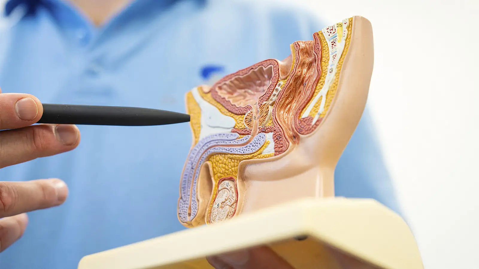
A CT scan showed a large, dense tube-like structure within the penile urethra, indicating a possible metallic core with surrounding calcification. There were no other urinary tract abnormalities.
The patient was started on intravenous piperacillin/tazobactam and clindamycin, and the team determined that endoscopic extraction of the foreign body wasn’t possible due to its size and significant edema. Instead, they inserted a suprapubic catheter to relieve urinary retention and divert the infected urine from the urethra.
Given the rarity of the presentation, clinicians discussed the case in a multidisciplinary andrology and genito-urethral surgery and radiology meeting, ultimately deciding that surgical removal should be delayed to allow time for the local infection and edema to improve with antibiotics. The patient was given a prolonged course of amoxicillin and discharged.
Four weeks later, the swelling had improved, and the patient was scheduled for open surgical removal 8 weeks later.
Under general anesthesia, the team performed a ventral urethrotomy and removed a 9 cm x 5 cm urethral stone. Within the core of the stone was “a degraded segment of a telephone cable.”
A transurethral catheter was left in place in addition to the suprapubic catheter. The patient was in good health after the operation and was discharged with a 2-week course of co-amoxiclav. Intraoperative lab tests confirmed the presence of E. coli and S. milleri.
The patient recovered from surgery without complications and with adequate wound healing. A follow-up visit showed a “satisfactory functional and cosmetic outcome” with no reported urinary symptoms, the authors reported.
“Self-inserted urethral foreign bodies are well described in the literature, spanning a wide range of objects such as cables, pens, wires, glass, or plastic items,” Bochinski and colleagues wrote. “Most patients, however, present within hours to days from insertion due to significant voiding symptoms and are therefore amenable to endoscopic removal.”
Boback Berookhim, MD, a urologist at Northwell Lenox Hill Hospital in New York City who was not involved in the case, said a key takeaway is the importance of making patients comfortable enough to share what is happening with them.
“It’s about having a nonjudgmental conversation,” Berookhim told MedPage Today. During his training, Berookhim and colleagues saw patients “who put foreign objects inside,” including some psychiatric patients with compulsions. However, he said, objects “retained for many years like this, I’ve never seen.”
Source link : https://www.medpagetoday.com/urology/urology/121314
Author :
Publish date : 2026-05-18 17:00:00
Copyright for syndicated content belongs to the linked Source.


